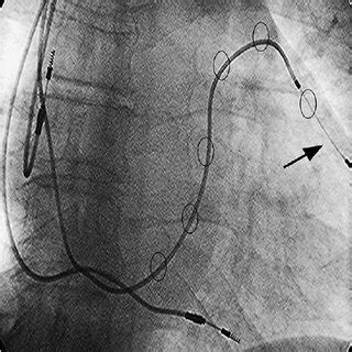
lv epicardial lead | epicardial lv lead replacement lv epicardial lead An optimal placement of the left ventricular (LV) lead appears crucial for the . LIV Golf Community - Join the conversation about the LIV Golf professional golf tour. Discuss events, teams, players, payouts, rankings, leaderboard and more here!
0 · transthoracic introduction epicardial lead
1 · transthoracic epicardial lead placement
2 · transthoracic epicardial lead insert
3 · left ventricular lead frontiers
4 · invasive epicardial lead placement
5 · epicardial lv lead replacement
6 · epicardial lv lead placement
7 · epicardial lv lead implantation
If you want to keep your pantry stocked with the best Filipino treats, you can never go wrong with Goldilocks! Check out our menu or visit our store to buy the best-baked delights in Sin City. 2797 South Maryland Parkway. Las Vegas, NV 89109. 702-368-2253.
Left ventricular (LV) lead positioning remains an important variable that predicts the response to CRT. Anatomical and technical challenges can hinder optimal LV lead placement using traditional lead implantation approaches.An optimal placement of the left ventricular (LV) lead appears crucial for the .
Between March 2008 and May 2014 an epicardial LV lead was implanted in 32 .

Epicardial LV lead positioning has the advantage of direct visualization and .Therefore, epicardial LV lead implantation using VATS can be effectively and .
An optimal placement of the left ventricular (LV) lead appears crucial for the intended hemodynamic and hence clinical improvement. A well-localized target area and tools that help . LV Lead Location and Baseline Clinical Characteristics. The LV lead position was assessed in 799 patients (55% patients ≥65 years of age, 26% female, 10% LVEF ≤25%, 55% ischemic cardiomyopathy, and 71% LBBB) .
transthoracic introduction epicardial lead
Minimally invasive left ventricular epicardial lead placement is safe and effective, offering selection of the best pacing site with minimal morbidity; it can be considered a primary option for resynchronization therapy.

Between March 2008 and May 2014 an epicardial LV lead was implanted in 32 patients after failed transvenous LV lead placement using a left-sided lateral minithoracotomy . The present article reviews the literature on image-guided cardiac resynchronization therapy (CRT) studies. Improved outcome to CRT has been associated with the placement of a left ventricular (LV) lead in the latest .
Epicardial LV lead positioning has the advantage of direct visualization and selection of the most suitable surface of LV, also avoiding areas of epicardial fat or fibrosis that .
Using an epicardial lead placed on the LV free wall via thoracotomy and endocardial leads placed in the right atrium (RA), left atrium (LA) via the coronary sinus (CS) .
transthoracic epicardial lead placement
Therefore, epicardial LV lead implantation using VATS can be effectively and safely used as a rescue method for patients with recurrent LV lead dislodgement, or even as a .
Left ventricular (LV) lead positioning remains an important variable that predicts the response to CRT. Anatomical and technical challenges can hinder optimal LV lead placement using traditional lead implantation approaches.An optimal placement of the left ventricular (LV) lead appears crucial for the intended hemodynamic and hence clinical improvement. A well-localized target area and tools that help to achieve successful lead implantation seem to be of utmost importance to .
LV Lead Location and Baseline Clinical Characteristics. The LV lead position was assessed in 799 patients (55% patients ≥65 years of age, 26% female, 10% LVEF ≤25%, 55% ischemic cardiomyopathy, and 71% LBBB) with a follow-up of 29±11 months.Minimally invasive left ventricular epicardial lead placement is safe and effective, offering selection of the best pacing site with minimal morbidity; it can be considered a primary option for resynchronization therapy.
We retrospectively assessed two types of sutureless screw-in left ventricular (LV) leads (steroid eluting vs. non-steroid eluting) in cardiac resynchronization therapy (CRT) implantation with regards to their electrical performance. Between March 2008 and May 2014 an epicardial LV lead was implanted in 32 patients after failed transvenous LV lead placement using a left-sided lateral minithoracotomy or video-assisted thoracoscopy (mean age 64 ± 9 years).
The present article reviews the literature on image-guided cardiac resynchronization therapy (CRT) studies. Improved outcome to CRT has been associated with the placement of a left ventricular (LV) lead in the latest activated segment free from scar. Epicardial LV lead positioning has the advantage of direct visualization and selection of the most suitable surface of LV, also avoiding areas of epicardial fat or fibrosis that can cause increase in pacing thresholds. Using an epicardial lead placed on the LV free wall via thoracotomy and endocardial leads placed in the right atrium (RA), left atrium (LA) via the coronary sinus (CS) and RV, they demonstrated a decrease in pulmonary capillary wedge pressure and an increase in cardiac output with temporary four-chamber pacing. Therefore, epicardial LV lead implantation using VATS can be effectively and safely used as a rescue method for patients with recurrent LV lead dislodgement, or even as a de novo way for those with unfavorable cardiac vein anatomy or when better targeted LV pacing is .
transthoracic epicardial lead insert
Left ventricular (LV) lead positioning remains an important variable that predicts the response to CRT. Anatomical and technical challenges can hinder optimal LV lead placement using traditional lead implantation approaches.An optimal placement of the left ventricular (LV) lead appears crucial for the intended hemodynamic and hence clinical improvement. A well-localized target area and tools that help to achieve successful lead implantation seem to be of utmost importance to .

LV Lead Location and Baseline Clinical Characteristics. The LV lead position was assessed in 799 patients (55% patients ≥65 years of age, 26% female, 10% LVEF ≤25%, 55% ischemic cardiomyopathy, and 71% LBBB) with a follow-up of 29±11 months.Minimally invasive left ventricular epicardial lead placement is safe and effective, offering selection of the best pacing site with minimal morbidity; it can be considered a primary option for resynchronization therapy. We retrospectively assessed two types of sutureless screw-in left ventricular (LV) leads (steroid eluting vs. non-steroid eluting) in cardiac resynchronization therapy (CRT) implantation with regards to their electrical performance. Between March 2008 and May 2014 an epicardial LV lead was implanted in 32 patients after failed transvenous LV lead placement using a left-sided lateral minithoracotomy or video-assisted thoracoscopy (mean age 64 ± 9 years).
The present article reviews the literature on image-guided cardiac resynchronization therapy (CRT) studies. Improved outcome to CRT has been associated with the placement of a left ventricular (LV) lead in the latest activated segment free from scar. Epicardial LV lead positioning has the advantage of direct visualization and selection of the most suitable surface of LV, also avoiding areas of epicardial fat or fibrosis that can cause increase in pacing thresholds. Using an epicardial lead placed on the LV free wall via thoracotomy and endocardial leads placed in the right atrium (RA), left atrium (LA) via the coronary sinus (CS) and RV, they demonstrated a decrease in pulmonary capillary wedge pressure and an increase in cardiac output with temporary four-chamber pacing.
rolex watch boxes to buy
rolex watch company story
Google asistents tagad ir integrēts pakalpojumā Google Maps, lai jūs varētu sūtīt ziņojumus, zvanīt, klausīties mūziku un izmantot brīvroku režīmu braukšanas laikā. Lai sāktu darbu, sakiet “Ok Google”. Jaunākā reāllaika informācija par sabiedrisko transportu.
lv epicardial lead|epicardial lv lead replacement


